Here you will find everything related to cephalometric analysis, digital radiography, Dental imaging basically everything ceph-related.

ORCA Dental AI Announces the Appointment of Mr. Chen Porat as VP Sales

  1. CephX | AI Driven Dental Services

ORCA Dental AI, the leading dental AI solutions provider, today announcedthe appointment of Mr. Chen Porat as VP Sales.

The move follows an extensive period of continuous growth and increasing market demand for the company’s new AI software products.

Mr. Porat joins us with over 8 years of sales management in the dental market, and extensive experience in working with key players and opinion leaders in the field.

In his previous position, Mr. Porat managed the US subsidiary of a leading dental implant company. In his role, he helped develop the company into a substantial market player by setting up a sales team, a marketing strategy, as well as an infrastructure that supported rapid growth.

As VP of sales, Mr. Porat will lead the ORCA Dental AI global sales team to establish a global network of resellers and agents and support continuous growth.

“Since the release of our latest dental AI software, we’ve been experiencing accelerated growth and increasing demand from potential resellers and agents that recognized the business potential we can deliver,” said Shlomi Avigdor, Co-founder and CEO of ORCA Dental AI. “Chen’s proven experience in dental sales management will help Orca execute its ambitious growth plans for 2020 and we’re thrilled to have him join the team.

About ORCA Dental AI

ORCA’s AI and deep learning solutions allow automated and accurate interpretations of dental imagery. The company’s technology helps dental practitioners to improve office efficiency and productivity, save time and energy, and to reduce medico-legal risk.

ORCA envisions a world where its highly sophisticated capabilities will immediately and seamlessly provide diagnostics, visual treatment suggestions and pathology findings. The solutions cover all types of dental imagery namely X-rays, CTs and intraoral scans.

ORCA aims to provide services to the entire dental ecosystem including Orthodontists, GPs and Prosthodontists. ORCA has partnered with the leading dental imaging manufacturers and top market players across the entire dental value chain, including Dentsply Sirona, Cefla, and Planmeca.

For more information about ORCA’s advanced solutions, visit http://www.orca-dental.ai.

Read more about How to Improve your Dental Practice
and Burstone Analysis

A Brilliant Way to Minimize Metal Artifacts in CBCT Scans

  1. CephX | AI Driven Dental Services

Artifacts in CBCT

CBCT technology is rapidly gaining popularity in the dental space, providing detailed and precise anatomical structure information and enabling advanced diagnostics and treatment planning for implantology, orthodontics, oral surgery, and endodontics.

CBCT technology has its limits, though, artifacts being a major one. Artifacts refer to variances between the reconstructed image and the actual content of the scanned subject. These variances usually make the results of scans diagnostically unusable as a result of their degraded quality. In addition to having degraded quality, structures that do not exist within the subject might appear in the images, creating a largely inaccurate scan result. These structures come about as a result of an imprecise image capture, reconstruction process or even patient motion. Artifacts make proper diagnosis very difficult and sometimes impossible to carry out. This article aims to highlight everything you need to know about artifacts and how to minimize them in CBCT scans.

Metal Artifacts

Metal streak artifacts are quite common in CBCT scans, often caused by patient-related objects such as implants, crowns, posts, fillings, and other radiopaque materials. These high-attenuation objects cause noise scattering and beam hardening, often resulting in bright and dark streaks that degrade the image quality.

Minimizing Artifacts in CBCT Scans

Minimizing metal artifacts produces a cleaner image, increasing diagnostics and improving treatment planning abilities. Other than the obvious, e.g. taking off metal objects (mainly jewelry), there’s another way to reduce artifacts – a brilliant one.

Deflection

Artifacts generated from metal or other radiopaque materials appear only in the plane horizontal to the patient positioning during the scan. By deflecting the artifact plane from the region of interest (ROI), you get a scan in which your ROI is cleaner, providing you with better diagnostics.

This deflection is executed by simply tilting the patient’s chin up or down when positioning for the scan.

For implant planning, the best patient positioning in most cases will be occlusal plane horizontal, leaving all crown and filling artifacts in the occlusal plane. In these cases, the bone will be as clean as possible.

It can still be affected by implants, deep posts, or gutta-percha.

When the ROI includes one or more teeth in the mandibular anterior region, position the patient with their chin tilted down. This way, artifacts generated in posterior areas will stay above the ROI.

ROI – mandibular posterior region: Place the patient with their chin tilted up. This will keep artifacts generated in the mandibular anterior region above the ROI.

ROI – maxillary anterior region: Place the patient with their chin tilted up. This will keep artifacts generated in the posterior area under the ROI.

ROI – maxillary posterior region: Place the patient with their chin tilted down. This will keep artifacts generated in the maxillary anterior region under the ROI.

ROI – premolars and first molars in either jaw: Place the patient with the occlusal plane horizontal. 

Make sure to help your patient place their heads in the various positions. Guide them in clear terms where to place their chin and change the machine height as needed.

In conclusion, it is easy to see that artifacts get in the way of carrying out proper diagnoses and treatment. Despite that, with careful planning and attention to detail, you can achieve good, usable results for your needs.


Read more about Sassouni Analysis
and Top Dental Products for 2015

5 Tools to Diagnose Impacted Teeth

  1. CephX | AI Driven Dental Services

5 Best Tools to Diagnose Impacted Teeth

Misdiagnosed impacted teeth may turn out to be risky to dentists just s they are to patients. Malpractice lawsuits are increasing in numbers, as patients claim for negligence- failure to timely recognize the need for eruption management, and failure to take timely action before risking the patients’ health.

The causes of an impacted tooth are often as a result of lack of space for the tooth in the mouth, due to orthodontic treatment or genetic factors. In other words, it may be caused by inherited factors or as a secondary effect to another oral condition.

This tooth condition can present with symptoms, i.e., symptomatic or even show no symptoms at all, i.e., asymptomatic. For those that present with symptoms, the common ones are:

  • Bad breath
  • Red swollen gum
  • Pain associated with mouth opening, biting, or chewing.
  • Bleeding gum etc.

The list of symptoms is not exclusive; some may experience one or more, while others none. And of course, these symptoms can serve as valid hallmarks to diagnosing the impacted tooth condition.

Here are some of the best methods of diagnosing impacted teeth:

  • Evaluation of the condition of tooth and gum: This is the conventional first diagnostic step taken by dentists. But this diagnosis is often not accurate, as there are many gums, teeth, or oral disease conditions that affect the gum, presenting almost the same symptoms as the “red- swollen- bleeding gum.” However, it can give a solid lead.
  • Intra-oral radiography – This type of radiography is said to more commonly used compared to the extra-oral radiography. The various Subtypes under it are indicative of the aspect of the teeth they show. Such Subtypes include:  Bite-wing x-rays, Periapical x-rays, Occlusal x-rays.

Known down-side for intra-oral radiography is their limited field-of-view, which can easily lead to missing out impacted teeth even when pointing the equipment to the right area.

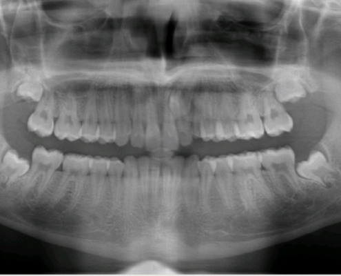
  • Extraoral radiography – Referring primarily to Panoramic x-rays, this is a method preferred by most dentists. It enables effectively detecting teeth impaction within a region of the mouth. This implies that the specificity of location of impacted teeth may not be ascertained.
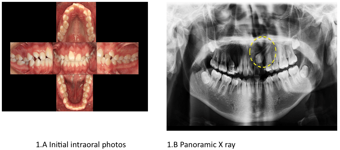
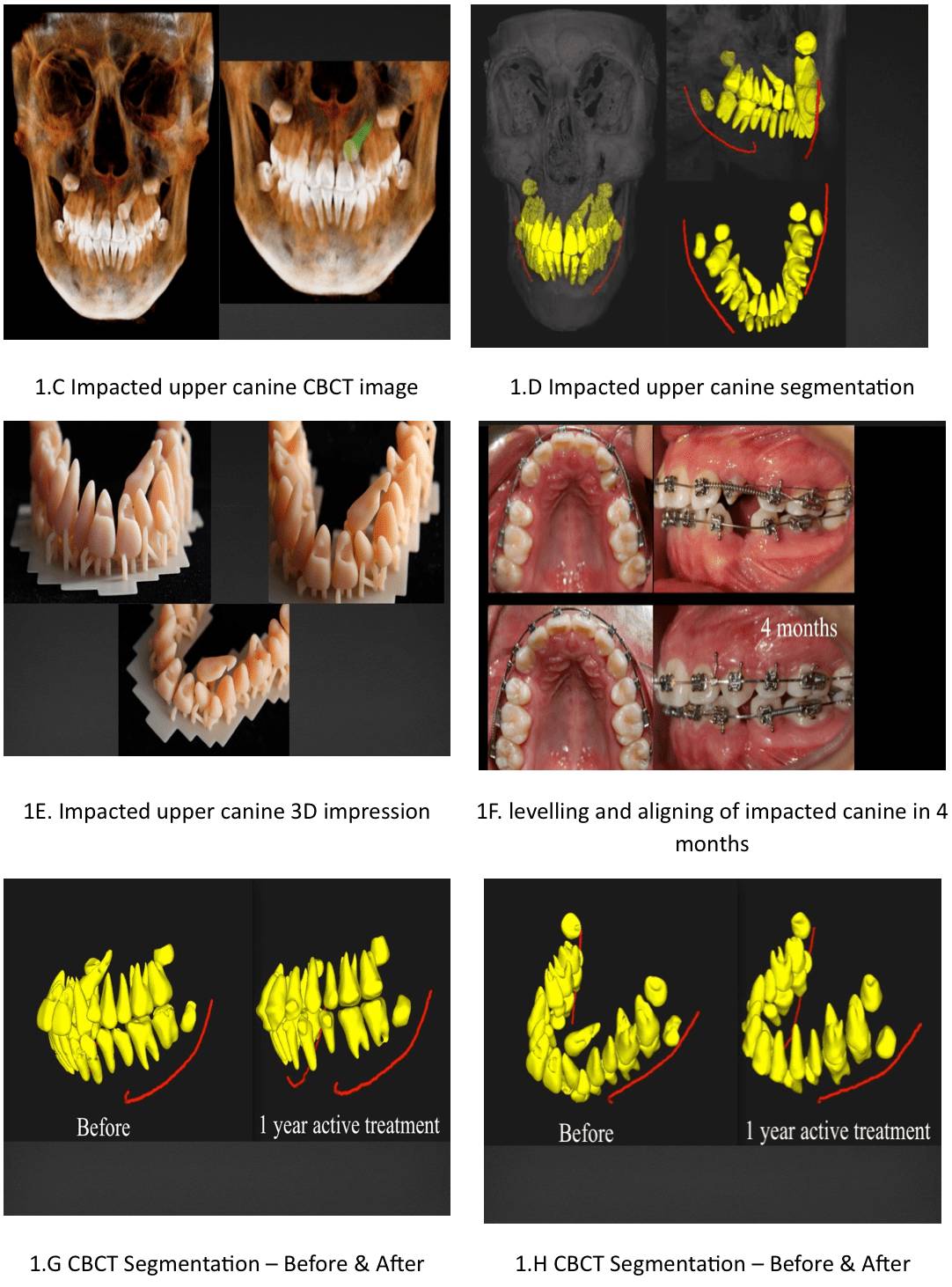
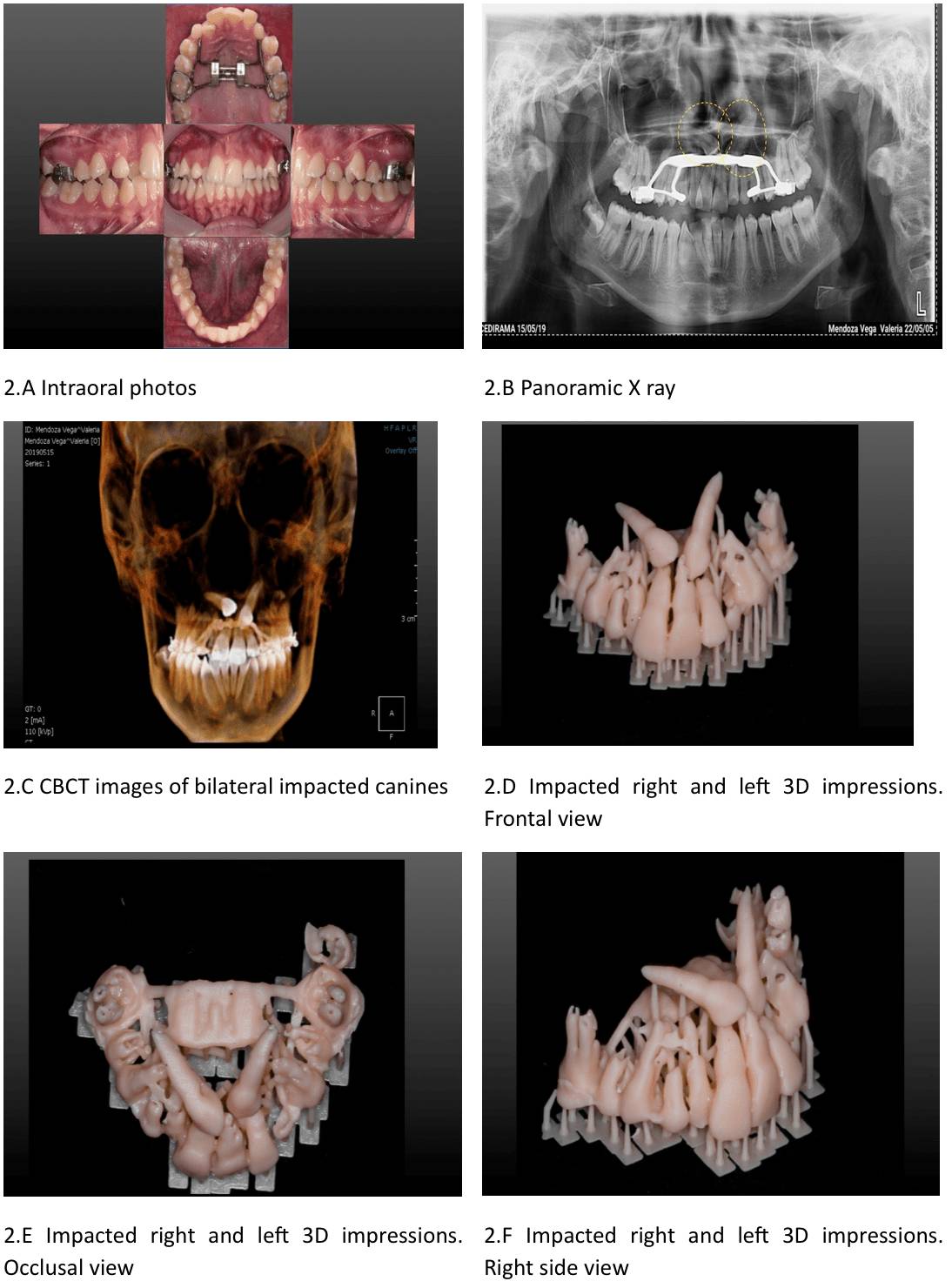
  • Cone-Beam Computed Tomography (CBCT):  Acquiring 3D scan is usually better, safer and more accurate than the radiography methods. For the safety of the patient it is often safer to take one exact CBCT in place of having to do repetitive radiography checks. The harmful effect of radiation is thus circumvented by this method of diagnosis. However many dentists find it hard to master 3D software, having to search through slices using different filters. Also, unlocking its full potential requires manual anatomy segmentation, before teeth and jaws can be moved, colored, removed etc, to allow diagnosis of impaction and other clinical condition, such as root absorption, bone thickness, nerve canal location and others.   
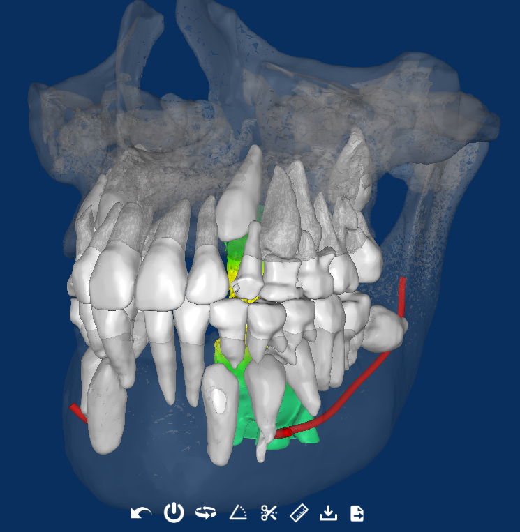
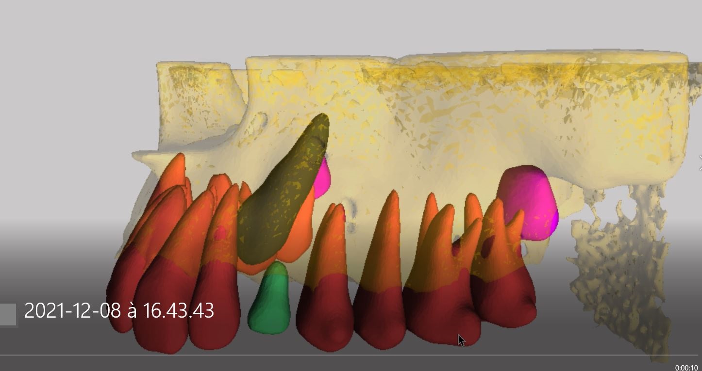
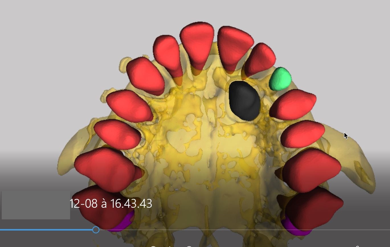
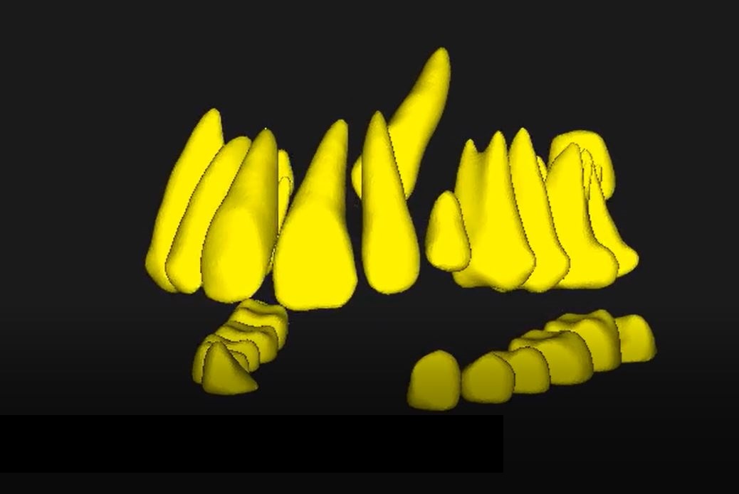
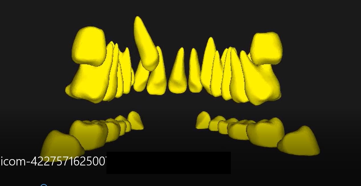
  • AI Driven 3D Segmentation: A 3D viewer that shows teeth and bone segmented by artificial-intelligence from a CBCT scan. This is an improvement over the conventional cone-beam computed tomography method of diagnosis, without adding any manual segmentation work or cost

Unlike the other methods of diagnosing impacted teeth, this newer method is better and more reliable. It gives a perfect 3D picture of the teeth segmented, making it very easy for the dentist/oral surgeon to accurately identify the impacted tooth/teeth. Dentists also have the option to share the case with patients, making them more connected and informed, to increase case acceptance.

CBCT 3D viewer helps the dentist to precisely identify the location of the impacted teeth; this is unlike the panoramic imaging that only helps to predict the teeth such as the maxillary canine. It reveals the location and shows the entire teeth in a three-dimensional manner, thus enabling accurate diagnosis.

Read more about How to Start an Orthodontic Practice
and Retainers and Maintenance: Preserving Your Patients’ Investment

Case Study – The Value of CephX Artificial-Intelligence (AI)

  1. CephX | AI Driven Dental Services
Read more about The Importance of Precise 3D Localization of Impacted Teeth Using CBCT in Orthodontics
and Unveiling Complex Orthodontics: How AI Transforms Diagnosis and Treatment Planning

What is Airway Volume Analysis?

  1. CephX | AI Driven Dental Services

Orthodontists have always had an interest in the upper airway, whether it’s the relationship between the airway and facial type, airway shape, or the airway volume. Most airway volume analysis is conducted using 2-dimensional (2D) cephalograms, which create a 2D airway analysis. This method provides limited data, like angular and linear measurements. A more sophisticated 3D airway analysis can be conducted by using 3D technology as the airway is part of a 3D structure. The most recent tool that can reveal the airway in 3D is cone-beam computed tomography (CBCT). The reason for orthodontists needing this information is so they can measure the airway volume for patients with obstructive sleep apnea, in relation to malocclusions, and also in those patients who have had orthodontic treatment.

2D lateral cephalograms

The older lateral cephalograms cost less than CBCT and are easy to use, but they are just limited to two-dimensional imaging, which doesn’t help to view structures in 3D. The use of CBCT has revealed that its reliability and accuracy of craniofacial measurements are far better than to 2D lateral cephalograms.

The use CBCT in the field of dentistry

This 3D diagnostic tool has in recent years become more accessible to the field of dentistry. It is now a reputable and well-acknowledged imaging technique for diagnosing oral and maxillofacial problems. It has in part become the preferred technique because it’s faster so it means the patient is subject to lower radiation doses. CBCT technology is so advanced that it can segment and visualise hollow structures in 3D such as airway volumes and surface areas.

Once the scan or digital image has been taken, the dentist, orthodontist or doctor needs to view the findings in a DICOM viewer, as DICOM is the file format accepted for a medical image. A DICOM viewer allows for a total analysis of any CBCT scan such as measuring airway volume and segmenting. When an image of an airway is segmented and structured this means delineating and removing any surrounding structures so that there is a clearer image which is easier to analyse.

Airway volume analysis and sleep apnea

The orthodontic community has a great interest in airway volume and respiratory function because studies have revealed airway problems are often related to different sorts of malocclusion and it’s been found that nasal obstruction is one of the main aetiological factors for dento-facial anomalies. Also, evaluation of airway volume is one of the diagnostic steps for patients who experience breathing disorders for example obstructive sleep apnoea (OSA) where the victims have marked craniofacial differences, like the position and size of the mandible, enlargement of the posterior airway space, and size of the soft palate and tongue. In these patients, airway volume analysis assessment has been performed, mostly using 2D lateral cephalograms, by identifying specific landmarks and measuring different areas and lengths in the airway region.

Airway volume analysis and malocclusion

Studies have shown that Class 11, division 1 malocclusion occurs because of an obstruction in the upper pharyngeal airway and mouth breathing. However, other research such as de Freitas et al. (2006), drew the conclusion that the width of the lower and upper pharyngeal airway width is not related to either Class I or Class II malocclusions. A further study by Kirjavainen and Kirjavainen in 2007 discovered that with Class II malocclusion, there is a definite association with the upper airway structure being narrower even in the absence of retrognathia.

Many of these studies to do with airway anatomy and its relationship to craniofacial development and growth face limitations because they have been using 2D frontal or lateral cephalograms which are not able to identify the contour of soft tissue in 3D which limits their ability to evaluate both volumes and areas in the upper airway. More and more practitioners take advantage of advances in computed tomography (CT) imaging and the 3D technology which allow them to better visualize the airway and volumetric analysis.

Clinicians are able to undertake the volumetric measurements and calculate the cross-sections of the airway in 3 planes of space, which are axial, coronal and sagittal. The axial plane, which can’t be seen on a lateral cephalogram, is considered to be the most relevant plane from a physiological perspective because it’s perpendicular to the airflow. CBCT systems have been specifically developed for the maxillofacial area.

Easy and accurate evaluation of the anatomy of the airway has been made possible using CBCT. Although there have been many studies published using CBCT to evaluate airways, not many have addressed its accuracy. Recent studies concentrated on evaluating both the reliability and accuracy of the airway volume which was measured on CBCTs digitally and at the most constricted part in the airway. This was compared to the manual measurements carried out on an airway model. They came up with varying results.

What we do at CephX

Our aim at CephX is to assist orthodontic practitioners worldwide to save valuable time by providing accurate analyses of dental imagery. The services we offer are automated, which is the highest quality analyses that helps you to improve the productivity and efficiency of your practice. Currently, we offer a cephalometric analysis service of 2D cephalometric x-rays or of CBCT scans, and 3D teeth segmentation. All the dental practitioner has to do is to upload the patient’s scan, and in no time at all he or she will be in receipt of the full analysis. Another key service we intend to add soon is automatic and instant 2D and 3D airway volume analysis using AI which will be available online.

Read more about Orthodontic Adverse Effects – Helping Patients Understand Them
and Build your Dental practice’s community reputation online